TOTAL KNEE REPLACEMENT
Orthopedic Surgery San Diego is a well-established orthopedic clinic in San Diego, CA delivering comprehensive orthopedic care to our patients in San Diego and surrounding areas. Our Total knee replacement service in San Diego is provided by expert surgeons, physiotherapists, and nurses. They’ll tailor a treatment and care plan to meet your needs and help you to get the most from your operation.
What is Total Knee Replacement?
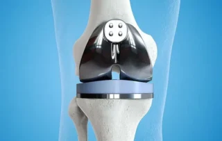
Total knee replacement, also known as total knee arthroplasty, is a surgical procedure in which a damaged or worn-out knee joint is replaced with an artificial joint or prosthesis. This procedure is commonly performed to relieve pain and restore function in individuals with severe arthritis or other conditions that affect the knee joint.TKA aims to improve the quality of life of individuals with end-stage osteoarthritis by reducing pain and increasing function and was found to improve patients’ sports and physical activity. The number of TKA surgeries has increased in developed countries, with younger patients receiving TKA.
Preoperative Optimization for Total Knee Replacement
Introduction
Adequate preoperative preparation and optimization is crucial for safe Total Knee Replacement or total knee arthroplasty (TKA) and good postoperative outcomes. Key elements include medical management, physical conditioning, patient education, and social support planning. Addressing modifiable risks preoperatively can reduce complications after Total Knee Replacement surgery.
Medical Risk Factor Optimization
Comorbid conditions like obesity, diabetes, hypertension, smoking, and anemia should be medically optimized. Losing weight, improving glycemic control, regulating blood pressure, smoking cessation, and iron supplementation all help reduce perioperative risks. Anticoagulation management is planned.
Nutritional Optimization
Protein intake and vitamin levels are screened and treated to help wound healing. Albumin levels predict outcomes. Malnutrition risks are addressed via diet, supplements, or referrals. Preoperative carbohydrate loading prepares metabolism for surgery and may reduce insulin resistance.
Physical Conditioning
Improving flexibility, muscle strength and endurance preoperatively enhances functional recovery. Aquatic therapy, home exercises, neuromuscular training, and gait aids expedite mobility gains after surgery. Stopping high-impact activities preoperatively also protects the joint.
Medication Management
Anti-inflammatory medications are typically held 1-2 weeks before surgery to reduce bleeding risks. Aspirin may be continued for cardiac patients. Reviewing supplements for their potential interactions is prudent. Pain medications are weaned appropriately.
Patient Education
Providing information on preoperative care, the procedure, rehabilitation stages, and expectations helps reduce anxiety while promoting participation in recovery. Clarifying restrictions, precautions, and follow-up needs ensures readiness.
Social Support Planning
Identifying family members or friends who can provide transportation to appointments, assistance at home, and emotional encouragement optimizes the support system. Preparing the post-discharge home environment also facilitates rehabilitation.
In summary, preoperative Total Knee Replacement surgery preparation encompasses medical, physical, educational, and social dimensions. Addressing modifiable risks and enlisting social support improves patient outcomes during the surgical episode of care. Thorough preoperative optimization is invaluable.
Surgical Steps for Total Knee Replacement
Introduction
Exposure
Femoral Preparation
Tibial Preparation
Patellar Resurfacing
Trialing and Final Components
Closure
The arthrotomy is closed, followed by deep dermal and skin closure. Local anesthetics are injected around the joint. Sterile dressings and compressive bandages are applied to complete the procedure.
In summary, precise bone cuts, soft tissue balancing, and methodical trialing allow accurate placement of TKA components in proper alignment for pain relief and restoration of function. Attention to each step optimizes clinical outcomes.
Robotic vs Traditional Total Knee Replacement
Introduction
Robotic systems have been introduced in Total Knee Replacement or total knee arthroplasty (TKA) to improve component position and surgical precision. Multiple randomized trials now allow the comparison of robotic-assisted Total Knee Replacement to conventional techniques.
Component Alignment
Robotic Total Knee Replacement results in fewer knee alignment outliers compared to traditional methods based on postoperative imaging. Robotic assistance reduces variance from desired alignment goals, with fewer knees falling outside the targeted coronal plane range. This could enhance implant survival.
Soft Tissue Balancing
Some robotic platforms quantify ligament tensions and gaps to help balance flexor and extensor mechanisms. This could optimize contact stresses and patellar tracking compared to traditional manual balancing. However, effects on functional outcomes are unclear.
Surgical Invasiveness
Existing robotic Total Knee Replacement systems require larger incisions for bone referencing and robot arm positioning. Increased surgical dissection could raise risks of pain and complications compared to minimally invasive surgery. Comparative injury rates are not yet well studied.
Functional Outcomes
Early data shows robotic Total Knee Replacement provides similar functional improvement and pain relief by 6-12 months postoperatively compared to conventional TKA.5 Range of motion and gait parameters are also comparable between techniques. Long-term monitoring is still needed.
Revision Rates
The impact of robotic TKA on implant survivorship and revision rates is not yet known, given its relatively recent development. Theoretically, improved position and balancing could reduce wear and failure. However, no long-term revision comparisons exist to date.
Cost-Effectiveness
Robotic systems involve high capital, disposable, and service costs. The charges add thousands of dollars per case compared to traditional Total Knee Replacement methods. It is unclear if the clinical benefits of robotics justify the added expense for hospitals and payers.
In summary, robotic guidance improves component positioning in Total Knee Replacement but long-term superiority over conventional techniques remains unproven. Comparative functional outcomes, complications, revision rates, and cost-effectiveness require further study to justify routine adoption of robotic platforms.
Postoperative Physical Therapy After Total Knee Replacement
Introduction
Early Postoperative Phase (0-2 weeks)
Intermediate Phase (2-6 weeks)
Advanced Phase (6-12 weeks)
Long-Term Considerations
Continued adherence to hip and core strengthening, cardiovascular exercise, and neuromuscular training helps maintain outcomes from Total Knee Replacement over years. Weight management optimizes joint loading. Preventing other joint problems promotes lifelong activity. Periodic follow-up helps track function. Most patients see benefits for 15-20 years postoperatively.
In summary, rehabilitation progresses through phases focused on mobility, strength, conditioning, and skills training. Patient participation is key for optimizing outcomes after Total Knee Replacement surgery. Physical therapy facilitates lasting functional gains.
Long-Term Infection Prevention After Total Knee Replacement
Introduction
Routine Health Maintenance
Dental Care
Prompt Infection Evaluation
Prudent Antibiotic Use
Skin Care
Careful cleansing and protection of open wounds, skin ulcers, or infections remote from the Total Knee Replacement prevent seeding deep infections. Small cuts should be monitored for signs of infection and treated promptly. Good hygiene is important.
In summary, maintaining wellness and immunity coupled with early infection detection optimizes prevention of late TKA infections. Patient education on precautions and prompt reporting of symptoms is key for successful long-term arthroplasty results.
Patient-Specific vs Kinematic Alignment in TKA
Introduction
Patient-Specific Instrumentation
Kinematic Alignment
Surgical Duration
Alignment Outcomes
Clinical Function
Conversion Rates
Kinematic alignment has a high rate of intraoperative crossover to conventional alignment when ligament releases fail to adequately balance the knee. Patient-specific guides function as cutting blocks so do not have conversion issues, but alignment may still be suboptimal.
In summary, both new techniques aim for more anatomically aligned TKA. Kinematic principles show early promise to improve function but remain challenging. Patient-specific guides have not proven superior despite higher costs. Long-term implant survival data is needed.
Functional Outcome After Total Knee Arthroplasty
Introduction
Baseline Function
Comorbidities
Preoperative Expectations
Postoperative Complications
Rehabilitation Compliance
Patients who actively participate in and adhere to postoperative rehabilitation have substantially higher functional gains from 6 weeks to 2 years after surgery. Compliance with exercise and mobility activity maximizes results.
In summary, patients with worse baseline function and fewer comorbidities tend to experience the greatest functional improvements from TKA. However, controlling expectations, avoiding complications, and commitment to rehabilitation play pivotal roles in optimizing individual outcomes.
RECOGNIZED BY