Knee Surgery, Knee pain treatment & Options in San Diego, CA


At Orthopedic Surgery San Diego, we offer superior minimally invasive options for every type of knee surgery, disease and symptom. Each knee surgery procedure we perform is supervised and administered by Dr. Robert Afra, whose longstanding experience at the forefront of orthopedic medicine has earned him a number of prestigious honors and patient recommendations. Dr. Afra provides cutting-edge surgical care including arthroscopic cartilage repair and ACL reconstruction, resurfacing techniques, partial knee replacement and total knee replacement. He places a premium on sound communication and lasting patient recovery, and works with you every step of the way to provide comprehensive, holistic treatment to restore your quality of life. If you’ve been told that you need knee surgery, we invite you to contact our knee surgery specialists to explore the various options at your disposal. We take pride in a patient-centered approach that addresses your questions and concerns throughout.

Knee Surgery San Diego

Our experience offers you practical ways to eliminate or reduce:
Knee injuries are some of the most debilitating events you can suffer. Whether you’ve torn the ligaments or broken a bone, the knee pain that accompanies such injuries can be paralyzing. Returning to everyday life and activities can be a challenge at best.

The knees are crucial joints that enable people to walk and perform other functions. Physical therapy often is effective in improving knee mobility and flexibility. In some cases, knee surgery is necessary to achieve long-term results. A range of surgical procedures can be employed to treat a variety of symptoms.  Your experienced San Diego orthopedic surgeon will walk you through every step of the process, from imaging and diagnosis to knee surgery and postoperative care. Dr. Robert Afra is widely recognized as one of the nation’s leaders in the field of knee surgery. Dr. Afra was the Chief of Sports Medicine in the UCSD Department of Orthopedic Surgery, and today runs one of the most respected knee surgery clinics in San Diego. With access to cutting edge technologies and an experienced technical team, Dr. Afra is proud to offer the finest orthopedic surgical care in Southern California. We have helped thousands of patients from across the country achieve lasting relief from knee injuries and symptoms. If you would like to schedule your own knee surgery consultation today, please contact us to learn more about the available options.


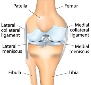
Knee Joint Description
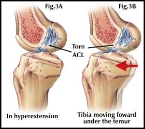
The human body is composed of many joints, such as the elbows, knees and hips. Every joint is important for movement and range of motion. The knee joint is where three bones join. They are the thigh bone, or femur; shin bone, or tibia; and kneecap, or patella. Four primary ligaments connecting the bones hold them together and maintain knee stability. Two medial and lateral collateral ligaments are located on the sides of the knee. They enable sideways movements of the joint. Cruciate ligaments, inside the knee joint, cross one other. The anterior cruciate ligament is in front, with the posterior cruciate ligament behind it. These ligaments are necessary to move the knee forward and backward. Two of the most common types of knee injuries are anterior cruciate ligament (ACL) sprains and tears, and posterior cruciate ligament (PCL) injuries.


Knee Surgery San Diego


ACL Tears


Because strenuous sports put a lot of stress on the knees, athletes often fall victim to ACL tears and sprains. About half of the time, patients with such conditions also suffer damage to other knee ligaments, the articular cartilage or the meniscus. Injured ligaments are considered “sprains” and are graded on a severity scale. A Grade 1 ACL sprain is the least severe, typically just an over-stretched ligament that does not impair normal knee function. A Grade 2 sprain involves a ligament stretched to the point that it is loose. This is sometimes called a “partial tear.” A complete tear, when the ligament is severed and the joint is unstable, is a Grade 3 sprain. According to the American Academy of Orthopaedic Surgeons, ACL injuries are caused by:

  1. Changing direction rapidly
  2. Stopping suddenly
  3. Slowing down while running
  4. Landing from a jump incorrectly
  5. Direct contact or collision, such as a football tackle.

Research indicates that women are particularly prone to ACL injuries. Certain sports, like football and basketball, are more of a threat to the knees than other physical activities. Symptoms include:

  1. A popping noise in the joint
  2. The knee giving out from under you
  3. Pain and swelling
  4. Loss of full range of motion
  5. Tenderness along the joint line
  6. Discomfort while walking.

A torn ACL does not heal on its own. Surgery is required to regain full function of the knee. Because the two sections of a severed ligament cannot be sutured back together, the ACL must be reconstructed by replacing it with a tissue graft on which a new ligament can grow. Graft material is taken from the patellar tendon, between the kneecap and the shin bone; the hamstring tendons, at the rear portion of the thigh; or the quadriceps tendon, which runs from the kneecap into the thigh. Surgeons sometimes graft tissue from cadavers. They perform the procedure by using an anthroscope to make small incisions. The operation involves less pain, shorter hospital stays and faster recovery times than more invasive surgery.
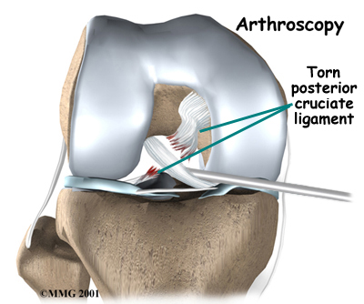


PCL Injuries


Because the posterior cruciate ligament is stronger than the ACL, it is more resilient to injury. The same three-stage severity scale used to evaluate ACL tears is employed to diagnose PCL injuries. It takes a powerful force to injure the PCL. Causes include: A direct blow to the front of the joint, like hitting the dashboard in a traffic accident or falling on a bent knee while playing sports; pulling or stretching the ligament, which can result in hyperextension; and stepping, walking or running in the wrong way. The symptoms of a PCL injury are:

  1. Pain in the back of the knee
  2. Swelling right after sustaining an injury
  3. Stiffness and difficulty walking
  4. A sense that the knee is about to give out.

In some cases, PCL injuries heal themselves and people regain full function of the knee. Physical therapy and nonsurgical procedures can be helpful. Knee Surgery is sometimes recommended, especially in the case of multiple injuries. For example, a patient might have a dislocated knee in addition to a PCL tear. As with the ACL, a torn PCL cannot be sewn back together. A tissue graft is required to rebuild it. Following knee surgery for an ACL or PCL injury, rehabilitation and recovery take six to 12 months. It is a challenging process, but the result is often a return to full function of the knee.


Knee Osteoarthritis


If your knee becomes inflamed due to arthritis, you will experience pain, swelling and stiffness. Left untreated, the discomfort and loss of mobility can progress and cause disability. While arthritis may occur in any joint, it is most often diagnosed in knees. There is no cure for arthritis, but treatment and knee surgery can relieve the symptoms and restore function. A layer of articular cartilage covers the three bones that connect in the knee, preventing them from rubbing against one another when the joint is flexed. Two other pieces of cartilage, called meniscus, form a cushion between the thighbone and shinbone. The synovial membrane, a lining around the joint, emits a lubricant into the cartilage. Osteoarthritis results from the gradual deterioration of the cartilage. Without this cushioning substance, inflammation and bone spurs can develop. This is the most commonly diagnosed type of knee arthritis. The condition worsens until it becomes difficult to bend or straighten the knee. The joint might buckle, lock in one position, make clicking or grinding noises, and get worse when the humidity rises. Other symptoms are redness, warmth and tenderness; and decreased range of motion. Medication, physical therapy and other methods can relieve some of the suffering. Another treatment, viscosupplementation, involves the injection of stem cells or platelet-rich plasma from your own body into the joint. Sometimes, knee surgery is necessary. Among the common procedures are:

  1. Arthroscopy, in which a surgeon makes a small incision and uses a thin instrument to diagnose and treat degenerated tissues;
  2. Cartilage grafting, the transplanting of healthy cartilage from another part of the knee or from a tissue bank;
  3. Osteotomy, in which a surgeon cuts and reshapes the shinbone or thighbone to take pressure off the joint; and
  4. Anthroplasty, the total or partial replacement of the knee with metal or plastic materials.

Knee Surgery


Knee Osteoarthritis Treatment Options


Analgesics/NSAIDS Use

Analgesics are medications that are taken to relieve pain. NSAIDS (non-steroidal anti-inflammatory drugs) like Motrin, are preferred over Tylenol, because of their antiinflammation effects; however NSAIDS are associated with more profound side effects. Tylenol has the tremendous downside risk factor of causing liver damage if taken in excess. While analgesics are not a cure for arthritis, they are generally used as a first line to treat osteoarthritis. In this stage of treatment, it is advisable to lower the degree of intense exercise and keep NSAID use to a minimum.

Injections

Short term pain relief can be facilitated by receiving cortisone or visco-supplementation injections. Cortisone, also known as methylprednisolone and triamcinolone, is a medication that can help relieve inflammation and pain in the joints. Pain relief can last from 3-6 months (occasionally more or less) from one injection, but it also carries a slight risk of infection. While this can relieve some pain for osteoarthritis sufferers, it is more effective for those with inflammatory arthritis due to the nature of the medication. Visco-supplementation, also known as synovial fluid replacement, Synvisc, and hyaluronic acid derivatives, is a series of 3 injections that are given one week apart. After the third injection, around 80% of patients experience some pain relief for an average of 8 months. It appears to work better during the early stages of osteoarthritis, and is less effective for those with other types of arthritis.

Topical Lotions

Diclofenac is an NSAID that when mixed with a solvent of dimethyl sulfoxide, or DMSO, creates a mild topical pain relief liquid. Although the topical cream does not work as effectively as the oral NSAID, the topical cream has much less significant side effect profile.

Dietary Supplements

Many patients claim to feel better from taking supplements, such as glucosamine, chondroitin, and MSM. It is recommended that a patient trial these supplements for at least one month prior to determining whether the supplement is effective in relieving pain.

Physical Interventions

Physical interventions are items applied to the skin to provide pain relief. Included are topical ointments, creams, magnets, heat packs, cold packs, pressure points, electrical currents, lasers, and exercise.

Exercise

While arthritis sufferers often shy away from exercise due to pain and inflammation, it is beneficial to keep the joint moving to maintain health and mobility. Weight gain, loss of muscle tone, or lack of motion can cause the arthritis to progress. Low-impact exercise such as swimming, walking, cycling, and even weight training may be possible if tolerated. Work with a trainer or physician, to find exercise that does not cause pain, but is personally enjoyable. Those with patella-femoral issues will benefit from quadriceps and core strengthening, as it relieves pain and helps increase range of motion. Weight should be maintained at the healthiest level possible with a BMI of around 25-29. Added weight puts more pressure on the joints. Pain can be lessened in many sufferers with only weight loss.

Unloader Braces

Medial, lateral, and patella-femoral unloader braces are available and have been found to relieve pain and decelerate the rate of degeneration. There are two type of braces for medial and lateral compartments: double-hinged, and single hinged. Double-hinged brace allows the skin to breathe a bit more than single-hinged, and is easier to wear under clothing. Custom-fit braces typically yield the best results.

Medial

Arthritis sufferers with damage in the medial compartment (the inner sides of the knee) can find pain relief while wearing a medial compartment brace. When worn in the early stages of osteoarthritis, the brace can slow down the rate of arthritis progression. Wearing a brace has also been shown to prevent surgery, and are used often for those with only a small part of the knee damaged, rather than the whole.

Lateral

Unloader braces have been shown to be beneficial for osteoarthritis sufferers in the lateral compartment arthritis (the outside of the knee). As with medial braces, using unloader braces early in the progression can slow down the degeneration in the area.

Patellar-Femoral

If suffering from patella-femoral osteoarthritis, unloader braces provide relief, especially to those whose damage is to the medial or lateral compartments. These braces allow for strength training. Unloader braces for this condition have more cushioning and are bulkier than the other types of braces.

Procedures:

Osteotomy is a surgical bone-cutting procedure used to lengthen, shorten, and/or reshape the bone. This procedure is used more often on relatively younger patients, who may be too young to undergo a partial knee replacement.

Opening Wedge Osteotomy of the Tibia (High Tibial Osteotomy)

The opening wedge osteotomy is used to realign bow legs, with the weight redirected to the lateral compartment that has been relatively well-preserved. This is commonly used for those with medial compartment osteoarthritis of the knee. In order for this procedure to be performed, the lateral compartment must 100% intact, the medial joint space must be at least 25-50% preserved, along with intact ligaments. During this surgery, the tibia is cut only once across. The bone is wedged open until the alignment is at its desired position. The space is kept open by using a plate or a block of bone. Healing time is about 6-8 weeks.

Opening Wedge, Distal Femoral Osteotomy

During this procedure, the femur (thigh bone) is cut above the knee in order to realign knock knees to the degree of bow legged. Essentially, the medial compartment of the knee is loaded to preserve the damaged lateral compartment of the knee.

Unicompartmental Knee Arthoplasty, Medial Mobile or Fixed Bearing

A medial compartmental knee replacement with a non-restrained Oxford knee (prosthesis) only replaces the part of the knee that is damaged. Between 90-95% of the prostheses have a positive outcome when an X-ray shows the lateral compartment is preserved, and an MRI shows the cruciate ligaments are functional. These can last from 10-15 years which is similar to the length of functionality of a complete knee replacement. A small percentage of patients will require a revision from a compartmental replacement to a total replacement, but it is an easier transition than going from one total replacement to a revision total replacement.

Total Knee Replacement

When osteoarthritis is too severe for non-surgical treatment, or a partial replacement, a total knee replacement is needed. The joint surface of the femur and tibia are replaced, with the replacement of the patella being a possible option. The percentage of positive outcomes are high with a 90-95% success rate 10 years after surgery, but varies individually based on lifestyle. Those who have a TKA due to an on the job (worker’s com) accident have a lower rate of about 40% for positive results. There are current developments in the surgery yielding smaller incisions, and less damage to surrounding tissue during the replacement. Total knee replacements should be avoided if a partial knee replacement can fix the issue. A partial replacement has lower risk of infection and more expeditious recovery. Knee replacements that acquire an infection can be disastrous with significant morbidity. A small percentage of patients, 5-10% need to have a new device within 10 years due to wear and tear. Total knee replacements are designed so that normal activities can be resumed, although running, jogging, and other high impact weight bearing activities can lead to replacement failure.

Arthroscopy for Osteoarthritis

Arthoscopy can temporarily help symptoms, such as knee locking or catching, known as “mechanical symptoms”, which are caused by loose cartilage flaps, meniscus tears, and rough joints. This outpatient surgery cleans up debris, smooths rough cartilage and meniscal damage which causes aches and pains during activities. The results from arthoscopy can last up to a few years, but varies per individual. Because the procedure shaves and smooths, it also wears down the cartilage, which will inevitably result in another procedure. Arthroscopy or lavage (flushing out the knee) for osteoarthritis symptoms that do not involve locking, catching, or giving way will likely not be helpful, although it can provide relief for a few months. Arthoscopy that is performed on a knee with non-mechanical symptoms are used for assessment, and is an alternative to an MRI. Many recent studies have shown that patients who undergo arthroscopy for osteoarthritis without mechanical symptoms are no better off than those patients who did not have surgery at one year out from the surgery.

Patellar Tendonitis

Patellar tendonitis affects a tendon of the same name, which connects the kneecap (patella) to the shinbone (tibia). This tendon helps you straighten your knee, jump and run. Sometimes called “jumper’s knee,” it often afflicts basketball and volleyball players. The initial signs of injury are pain and inflammation between the kneecap and the place where the tendon attaches to the shinbone. This is an overuse condition that gets worse when you exercise or perform repetitive motions. If medication, therapy, corticosteroid injections and other techniques fail, surgery could be warranted. The minimally invasive procedures entail only small incisions around the knee.

Quadriceps Tendonitis

Technically called tendonopathy, quadriceps tendonitis is another overuse injury frequently suffered by athletes. It features tissue damage and inflammation in the tendon that connects the quadriceps with the top of the kneecap. This causes pain in the front part of the knee, just above the kneecap. The quadriceps is essential for straightening and bending the knee. It cannot function properly if the tendon is damaged due to repetitive or prolonged activities. Symptoms include pain, swelling, stiffness and weakness in the joint. Most people suffering from this injury find relief with medication, physiotherapy and activity modification. Some receive injections of platelet-rich plasma. A common surgical approach is to remove degenerative tissue and calcifications from the tendon. In some cases, the tendon needs to be reattached to the kneecap.

Pes Bursitis


Inflammation of the bursa, between the shinbone and the hamstring muscle tendons, is called pes anserine bursitis. Bursae are small sacs containing a fluid that cushions joints. Inflammation results when the bursa produces excess fluid and causes swelling. The main symptoms are pain and tenderness on the inside of the knee, two or three inches below the join. The condition occurs due to overuse, or repetitive friction irritating the bursa. Runners and other athletes are especially vulnerable. Among the causes of pes bursitis are:

  1. Improper training methods, like failing to stretch
  2. Excessive running on hills, or a sudden increase in running distance;
  3. Tight hamstring muscles
  4. Obesity
  5. Turning out the knee or lower leg;
  6. Osteoarthritis in the knee
  7. A medial meniscus tear.

The first remedies to try are rest, ice treatments, anti-inflammatory medication and physical therapy. In advanced cases, knee surgery involving surgical removal of the bursa is necessary. The procedure frequently is performed on an outpatient basis.


Shin Splints


The common term for pain along the inner edge of the shinbone is shin splints. The condition usually develops following exercise, with runners and dancers among those most at risk. Technically known as medial tibial stress syndrome, shin splints are characterized by inflammation of the muscles, tendons and bone tissue around the shinbone. The pain occurs where the muscles connect to the bone. Causes of shin splints include:

  1. Strenuous physical activity
  2. Sudden changes in workout routines
  3. Flat feet or rigid arches
  4. Exercising with inadequate shoes.

Rest, ice, medication, stretching exercises, orthotics and elastic compression bandages often relieve the symptoms. Knee Surgery to remove the fascia (tissue around the muscles) could be needed if a patient’s condition involves compartment syndrome, or if the pain is extreme. When a muscle has been torn away from the shinbone, knee surgery is necessary to reattach the muscle.


Osteochondral Cartilage Lesion

When knee cartilage is damaged, bones in the joint are left unprotected. A common diagnosis for this condition is osteochondral cartilage lesions, which can involve cartilage tears, fractures or cysts. The damage occurs between the thigh bone (femur) and shin bone (tibia).

Cartilage Lesions result from:

1. An injury to the joint caused by twisting 2. The strain of playing football, golf and other sports 3. Inherited susceptibility to the condition 4. Improper bone development 5. Repetitive stress on the joint

Symptoms of the condition include:

1. Knee pain that does not subside following an injury 2. A persistent, mild ache in the joint 3. Knee locking or clicking 4. A loose feeling in the joint Doctors often initially recommend braces and casts. Usually, however, surgery becomes necessary. The primary procedures are: 1. Arthroscopic treatment, to extract damaged cartilage 2. Drilling into the bone to cause bleeding, which promotes healing 3. A bone graft to replace damaged cartilage 4. Knee replacement

Meniscus Tear

A type of cartilage called meniscus cushions and supports the thigh and shin bones. Meniscus tears often occur as a result of strenuous athletic activity, severe twisting of the knee, squatting or blunt-force injury. Cartilage gets thinner and weaker with age, making older people more vulnerable. Names for the various types of meniscus tears include longitudinal, parrot-beak, flap and bucket handle. The symptoms of a meniscus tear are: 1. A popping sound when the injury occurs 2. Pain and swelling 3. Stiffness and inflammation that worsens 4. Locking of the knee 5. Joint slipping or “giving out” 6. Decreased range of motion The type and size of a meniscus tear, as well as its location, determine the treatment. Doctors prescribe medication and the formula of rest, ice, compression and elevation (RICE) for sudden tears caused by injuries. A longitudinal tear, which occurs in the outer part of the cartilage, sometimes heals itself. A tear in the inner meniscus, where there is less blood to provide the nutrients necessary for healing, usually requires surgery to correct. Procedures include: 1. Arthroscopy, the insertion of miniature instruments to repair cartilage 2. Meniscectomy, the trimming of torn meniscus 3. Meniscus repair, stitching together torn pieces of cartilage

Patellofemoral Syndrome

Athletes and others who place excessive strain on their knees suffer pain classified as patellofemoral syndrome. Most at risk are those involved in activities like running, jumping and cycling. The syndrome is one of several conditions collectively called “runner’s knee.” Causes of the syndrome include: 1. Improper kneecap alignment 2. Total or partial joint dislocation 3. Overuse or injury 4. Excessive weight 5. Weak or imbalanced muscles 6. Inadequate stretching before exercise 7. Flat feet 8. Tissue irritation in the front of the joint 9. Strained tendons The main symptom is an ache in the patella (the front of the kneecap), at the point it meets the femur (thigh bone). Pain results from activities that entail flexing the knee and putting pressure on it. Sitting for long periods with the knees bent also causes discomfort. Nonsurgical treatments, such as anti-inflammatory drugs and the RICE formula, are sufficient in many cases. Exercising, taping the kneecap, bracing the joint and wearing orthotics can aid in recovery. Surgical solutions are arthroscopy, in which damaged cartilage is removed; and kneecap realignment, to reduce pressure on the cartilage.

Patellofemoral Dislocation

The patella connects the kneecap and thigh bone. It slides up and down when the knee is flexed. The patella can become completely or partially dislocated when an injury forces it to move sideways. Sometimes, it slips back into place, but not without significant pain and temporary loss of function. The typical cause of these dislocations is blunt-force trauma, like a fall or a blow to the knee. They also can result from straining or twisting the joint. In some cases, the groove in the femur through which the patella slides is not deep enough. That increases the odds of the patella slipping out of place. People with loose ligaments and overly flexible joints also are at risk. Symptoms of patellofemoral dislocation are: 1. A popping sound when an injury occurs 2. Pain and swelling in the kneecap 3. Feeling the kneecap shift out of place 4. The buckling or “giving out” of the joint 5. Deformity of the knee’s shape Doctors generally can manually move a patella back into place by employing a technique called reduction. A patient might wear a brace, or use crutches, for a few weeks to allow the knee to heal and strengthen. Physical therapy is often necessary. Due to tissue damage caused by dislocation, the patella is looser and liable to slip again. When that happens repeatedly, and the knee remains unstable, surgical alternatives are available. They generally involve arthroscopically reconstructing ligaments that support the patella. More invasive surgery is necessary when continual dislocation is a result of a bone deformity or hereditary condition.


RECOGNIZED BY


© 2023 Dr. Robert Afra – San Diego Orthopedic Surgery Shoulder – Knee – Elbow